Tuesday 10 July 2012

The Use of Donor Lymphocyte Infusion’s (DLI’s) and Early Treatment for Relapse

Presented on Friday, 29 June 2012

Introduction to Bone Marrow Transplant Nursing

Aim & Objectives

Aim: to gain a basic understanding of what DLI’s are and their use in BMT.
Objectives:
• Define DLI’s
• Discuss where they fit in the treatment algorithm of BMT

Definition of DLI

• Donor Lymphocytes Infusions
• Also known as Leukocyte infusions or Buffy coat infusions
• Cytotoxic T Lymphocytes
• DLI may have modified cells à T helper, Cytokines (IL-2, GM-CSF, Interferon)

Lymphocytes

• B Cell
• T Cell à
- Helper (CD4+), to promote other cells function
# T Helper 0 à
# T Reg
# T helper 17
- Cytotoxic (CD8+) à Effector cell
- Natural Killer Cell à Innate immunity
- Gamma delta
- Memory T cell à Helper & Cytotoxic

Minimising Toxicity

• Minimise toxicity à reduce Transplant Related Mortality (TRM) à Relapse
• Relapse rates are higher in patient who don’t develop GVHD

Maximising Toxicity

• Maximise anti tumour effect à High morbidity and mortality or High Transplant Related Mortality (TRM)
• Relapse rates are lower in patient who develop GVHD versus who don’t
• Same cell that cause GVHD give some tumour effect therefore protection à Disease not return

Balancing

• Allogeneic HSCT
• Combination of high dose chemotherapy & Allogeneic transplant à Remission
• Disease relapse is a major cause of treatment failure and salvage treatment options for these patients are limited

Relapse After Allogeneic HSCT

• No further therapy
• Withdrawal of immunosuppression to elicit a therapeutic graft-versus-tumor (GVT) response
• Reinduction chemotherapy with the same or different agents
• Or second transplants with or without preceding chemotherapy à Risk of serious, life-threatening complications
• Donor lymphocyte infusions with or without preceding chemotherapy à eliminate the need for second transplant.

Background

• Patient who received syngeneic stem cell transplants relapsed more frequently than those who received allogeneic transplant.
• A rapid decrease or elimination of post grafting prophylaxis or treatment immunosuppression would often lead into remissions.
• Higher numbers of relapses were seen in patient who received T Cell depleted grafts compared to patients who received unmanipulated grafts.

Withdrawal of Immunosuppression

• Cyclosporine à blocks interleukin-2 (IL-2) transcription on the T cell
• Tacrolimus à inhibit IL-2 synthesis
• Graft versus Leukemia effect
• HSCT was used to allow for higher doses of chemotherapy and radiation therapy during treatment
• Graft-versus-leukemia/lymphoma (GVL) effect àin which the new immune cells produced by a matched donor (allogeneic) transplant can recognize cancer cells as a threat to the body and destroy them, reducing the rate of disease relapse.

Collection of DLI

• Collected from original stem cell transplant donor
• Obtained by using automated Apheresis equipment
• Collected cells may be infused unmanipulated or may be manipulated

The Goal of Therapy

• The goal of this therapy is to induce a remission of the patient's cancer by a process called the graft versus tumour effect (GVT). The donor T-cells can attack and control the growth of residual cancer cells providing the GVT effect.
• It is hoped that the donor leukocyte infusion will cause GVT and lead to a remission of the patients cancer.
• Patients might require standard chemotherapy, to reduce the amount of cancer cells they have prior to their donor lymphocyte infusion.

Cell Dose

• Ranged from 0.1 to 8.8 x 10⁸ T Cell per Kg.
• @ RPAH we collect T Cell (CD4 positive T cell) ranged from 1 x 10⁷ to 5 x 10⁷ per Kg.
• Divided according to request
• How many T cells should be infused in the dose of DLI for prophylaxis or treatment?
• How many doses should be given?
• How frequently they should be given to maintain a GVL effect?
• When should infusion be stopped?

Un-manipulated Lymphocytes

• Harvested and infused fresh
• Harvested & cryopreserved
• Harvested, selected & infused fresh
• Harvested, selected & cryopreserved
• To prevent or treat relapse of the disease
• To treat mixed or decreasing chimerism
• To treat infections
• Prophylaxis or pre emptive therapy for relapse, chimerism or infections

Manipulated Lymphocytes

• Primed with expanded with cytokines
• Cultured with antigens, viruses, etc
• Gene modified with suicide genes
• Depleted of unwanted lymphocytes
• To enhance immune reconstitution
• To improve engraftment
• To prevent or modulate GvHD
• In combination with other cells such as mesenchimal stem cells, dendritic cells, etc

Response to DLI post Allo Tx.

• CML > 70%
• MM : 40%
• NHL : 20%
• Acute Leukemias : 15%
à MDS 36%, AML 22% & ALL 8%

Assessing efficacy of DLI:

- Morphologic response
- Molecular response
such as BCR-ABL for CML

CML

• DLI is best suited for treating slowly relapsing Leukemia
• The use of DLI to boost anti leukemia/anti lymphoma effects in patient who fail develop GvHD
• The use of DLI for patients with only cytogenetic/molecular relapse continues to give excellent remissions rates

AML

• DLI for relapse AML following HSCT is not very effective in getting patient into remission.
• Lower initial disease burden, reductions in the tumour burden with chemotherapy prior to DLI and favourable cytogenetic appear to be beneficial for patient with relapse leukemia after SCT

ALL

• Responds even more poorly than AML
• The poor response of relapse to DLI is thought to be mediated by diverse immune escape mechanisms of leukaemia cells

Lymphomas

• Highest response rates were seen in the indolent lymphoma
• More aggressive lymphomas had a lower response rates
• Lower tumour burdens and slower growing malignancies are essential conditions for DLI to be effective

Multiple Myeloma

• The role of DLI is unclear, but can be considered

Complications

• Complications of DLI include acute and chronic graft versus host disease and bone marrow aplasia
• Resulting in immunosuppressant and susceptibility to opportunistic infections.
• Progessive disease is the major cause of death after DLI followed by GvHD, infections and pancytopaenia

RPA Experience

• 17 à Donor Lymphocyte collected from 2001 to 2011
• 17 à Patients had Donor Lymphocyte Infusions from 2001 to 2011

Summary & Future Direction

• Donor lymphocyte infusion (DLI) attempts to stimulate a donor versus leukemic reaction and thus eradicate the malignant clone of cells. in hopes of triggering additional GVL activity.
• But this option is effective only in a minority of patients, mostly those with chronic myelogenous leukemia.
• Searching for ways to increase the effectiveness of donor leukocyte infusions and minimize its side effects.

References

• Campregher, PV, Gooley, T, Scott, BL, Moravec, C, Sandmaier, B, Martin, PJ, Deeg, HJ, Warren, EH & Flowers, MED 2007, Result of donor lymphocyte infusions for relapsed myelodysplastic syndrome after hematopoietic cell transplantation, Bone Marrow Transplantation, 40, pp. 965-971
• Deol, A & Lum, LG 2010, Role of donor lymphocyte infusions in relapsed hematological malignancies after stem cell transplantation revisited, Cancer Treatment Reviews, 36, pp. 528-538.
• Luznik, L & Fuchs, E 2002, Donor lymphocyte infusions to treat hematologic malignancies in relapse after allegeneic Blood or marrow transplantation, Cancer Control, 9 (2), pp. 123-137.
• Roush, KS & Hillyer, CD 2002, Donor lymphocyte infusion therapy, Transfusion Medicine Reviews, 16 (2), pp. 161-176.

Friday 31 December 2010

Leukemia Kronik

LEUKEMIA KRONIK


ETIOLOGI
Penyebab penyakit tidak diketahui secara pasti. Sama seperti tipe leukemia yang lainnya, leukemia berasal dari mutasi yang terjadi pada spesifik protein yang disebut juga dengan gen yang mengkontrol perkembangan dan pertumbuhan dari sel darah. Akibatnya sel berkembang dan bertumbuh tidak terkontrol.


LEUKEMIA LIMFOBLASTIK KRONIK


Leukemia limfoblastik leukemia (LLK) adalah gangguan pada monoklonal yang dikarakteristikkan dengan adanya proliferasi limfosit B meskipun proliferasi dari limfosit T terjadi tetapi sangat jarang. Pada leukemia ini limfosit diproduksi tetapi tidak berfungsi, dan berakumulasi di dalam darah, sumsum tulang dan jaringan limfa. Setiap individu berbeda dalam jumlah akumulasi. Biasanya leukemia ini banyak terjadi di negara-negara eropa. Jenis lainnya yang dapat dimasukkan ke dalam kategori LLK adalah sindroma sezary (merupakan fusi leukemia dari mikosis fungoides) dan leukemia sel berambut (menghasilkan sejumlah besar sel darah putih yang memiliki tonjolan khas seperti rambut bila dilihat dibawah mikroskop).

GAMBARAN KLINIS

Leukemia limfoblastik kronik mempunyai bermacam-macam jenis. Banyak pasien yang tidak menunjukkan gejala menderita LLK. Gambaran klinis diantaranya letih lesu, infeksi, hilangnya berat badan, keringat malam, limfadenopati dan hepatosplenomegali. Gambaran klinis yang lain diantaranya adalah Infeksi, fatigue, limfadenopati (80%), hepatomegali/splenomegali (50%). LLK biasanya dimulai dengan limfositosis diikuti dengan limfadenopati dan kegagalan pada sumsum tulang, 10% adanya Coomb test. Adanya limfositosis adalah merupakan ciri khas dari LLK. Biasanya 75% hingga 98% sel yang beredar didalam darah adalah limfosit. Jumlah limfosit lebih dari 5000/mm3. Bentuk dari limfosit biasanya kecil dan sudah dewasa. Jumlah sel darah putih meningkat lebih dari 20.000/mm3. Sedangkan jumlah hematokrit dan trombosit biasanya normal. Lebih dari 30% limfosit menginfiltrasi sumsum tulang.

STADIUM DAN KLASIFIKASI

Menegakkan klasifikasi atau fase dari LLK sangat penting karena berguna dalam pengambilan keputusan apakah harus diterapi atau tidak serta dalam menegakkan prognosa. Ada 2 sistem yang dipergunakan dalam pengklasifikasian stadium LLK yang berdasarkan karakteristik dari sel yaitu klasifikasi RAI dan BINET. Klasifikasi RAI lebih populer dipergunakan di Amerika, klasifikasi ini mengkategorikan LLK menjadi resiko rendah, sedang dan tinggi. Sedangkan Klasifikasi Binet lebih populer di Eropa, klasifikasi ini berdasarkan jumlah jaringan limfosit yang terganggu, anemia dan trombositopenia. Klasifikasi ini sangat simpel dan berkorelasi dengan survival.

Klasifikasi RAI

Stadium Penemuan Survival

0 Hanya limfositosis
>120 bulan
I Limfositosis plus limfodenopati
95 bulan
II Limfositosis plus spleenomegali atau hepatomegali atau keduanya
72 bulan
III
Limfositosis plus anemia 30 bulan
IV
Limfositosis plus trombositopenia 30 bulan


Klasifikasi BINET

Stadium Penemuan Survival

A Hb> 10, trombosit >100 , <3 area yang terpengaruh
>120 bulan
B Hb> 10, trombosit >100 , >3 area yang terpengaruh
84 bulan
C Hb< 10, trombosit <100 , <3 area yang terpengaruh termasuk cervical, nodul axila atau inguinal, spleen atau hati.
24 bulan


DIAGNOSA

Diagnosa ditegakkan berdasarkan pemeriksaan darah biasanya ditemukan secara tidak sengaja dengan hitung limfosit yang sangat tinggi atau disebut juga limfositosis. Menurut Mulligan (2008), penegakkan diagnosa dapat disimpulkan dari fitur pemeriksaan klinis dan fitur pemeriksaan laboratorium.

I. Fitur Pemeriksaan Klinis.
Ditemukan adanya limfadenopati dan hepatosplenomegali yang mengakibatkan infeksi, autoimunitas dan transformasi.

II. Fitur Pemeriksaan Laboratorium.
a. Morfologi – pada pemeriksaan hitung darah lengkap mungkin ditemukan adanya abnormalitas pada ukuran dan bentuk dari sel darah. Hitung sel darah merah, sel darah putih dan trombosit juga menunjukkan adanya perbedaan. Trombositopenia dan anemia biasanya terjadi pada penyakit yang sudah lanjut. Lebih dari 90% bentuk dari limfosit berukuran kecil atau sedang.
b. Fenotipe – adalah test yang dipergunakan untuk mendeteksi antigen yang ditemukan pada permukaan disekitar sel (surface). Antigen sering diidentifikasikan sebagai Cluster Differentiation (CD), diikuti oleh nomer. Contohnya adalah CD19, CD20, CD5 dan CD23. Test ini sebaiknya dilakukan incase jumlah limfosit rendah pada waktu penegakkan diagnosa LLK. Imunofenotipe menunjukkan adanya limfosit B, CD19, CD20 dan CD5.
c. Pemeriksaan sumsum tulang – pemeriksaan sumsum tulang dengan biopsi yang mengkonfirmasikan diagnosa dari LLK. Translokasi jarang terjadi, tetapi yang lebih sering terjadi adalah delesi. Kromosom yang terganggu adalah 13q (berkisar 55%), 11q (18%), trisomi 12q (16%), 17p (7%), 6q (6%) dan normal sitogenetik sekitar 18%. Delesi kromosom 11q dan 17p berhubungan dengan prognosa yang buruk dan berkembang pesat. Sedangkan delesi kromosom 13q berhubungan dengan prognosa yang baik serta survival yang lama. Prognostik Marker CD38, menunjukkan LLK yang agresif.

Pemeriksaan lain yang dibutuhkan dan cukup bermanfaat adalah:
1. Direct Antiglobulin Test (DAT) – pemeriksaan ini sangat penting pada semua pasien anemik dan sebelum terapi dilaksanakan.
2. Hitung Retikulosit
3. Fungsi ginjal dan hati
4. Serum protein elektroporesis – adanya peningkatan. Tingkat serum imunoglobulin dalam darah – Hipogammaglobulinemia, hampir setengah populasi menderita, pada stadium lanjut hampir semua pasien menderita. Bila seseorang tingkat antibodinya rendah, orang tersebut dapat lebih mudah menderita sakit. Oleh karena itu transfusi imunoglobulin mungkin bermanfaat bagi pasien. Adanya mutasi pada imunoglobin rantai berat (heavy chain) - prognosa buruk.
5. Meningkatnya jumlah limfosit 2 kali lipat dalam satu tahun mempunyai surival yang lebih rendah dibandingkan dengan meningkatnya limfosit 2 kali lipat lebih dari 1 tahun
6. Pemeriksaan Xray pada paru-paru
7. Biopsi nodul limfa – dalam beberapa kasus pemeriksaan biopsi pada limfa mungkin dibutuhkan untuk membedakannya dengan limfoma atau bila dicurigai telah bertransformasi ke limfoma.
8. CT (Computerisec Axial Tomography) Scan – berguna bila dicurigai adanya splenomegali pada pemeriksaan fisik untuk mengkonfirmasikannya.
PROGNOSA

Prognosa tergantung dari area yang terpengaruh serta hasil pemeriksaan sel darah merah dan trombosit. Stadium A, merupakan stadium awal  tidak memperlihatkan gejala, nil terapi. Stadium B dan C membutuhkan terapi. Sepertiga dari LLK tidak membutuhkan terapi dan dapat hidup lebih dari 10 tahun, sedangkan sepertiga lagi berproses ke stadium berikutnya dan membutuhkan terapi, sisanya agresif dan membutuhkan terapi setelah didiagnosa. Tidak ada terapi dapat menyembuhkan LLK. Tujuan utama adalah mengkontrol simptom dan kemungkinan memperpanjang usia. Berkisar 5% Penderita LLK berkembang menjadi Large Cell Lymphoma yang sangat agresif. Prognosa pada pasien ini buruk dan hanya 5 bulan survival.


Baik Buruk
Jenis Kelamin
Perempuan Laki-laki
Stadium
A B, C
Limfosit waktu doubling
> 1 tahun < 6 bulan
ZAP 70
Negative Positive
Mutasi Somatik
Mutasi Germline
Sitogenetika
13q delesi Trisomi 12, delesi p53


PENATALAKSANAAN LEUKEMIA LIMFOSITIK KRONIK

Terapi pada Leukemia Limfositik Kronik tergantung dari stadium dari penyakitnya, mulai dari hanya mengobservasi hingga kemoterapi dan transplantasi sumsum tulang. Pada stadium awal (RAI I & II serta BINET A) tindakan hanya mengobservasi. Terapi dimulai ketika pasien mulai mengalami simptom dari penyakitnya. Pada stadium lanjut (RAI III & IV serta BINET A & B) dibutuhkan terapi. Terapi termaksud kemoterapi baik single maupun kombinasi, monoklonal antibodi, transplantasi sumsum tulang dan dosis rendah radioterapi. Pada pasien ini imun sistem mungkin terganggu, oleh karena itu antibiotik dan anti jamur mungkin dibutuhkan. Selain itu penggunaan immunoglobulin juga diberikan untuk meningkatkan sistem imunitas dan mencegah infeksi.

Chlorambucil(Leukeran)
Adalah kemoterapi oral jenis alkylating yang mana dipergunakan lebih dari 40 tahun dalam mengobati LLK.

Fludarabine
Adalah terapi standar yang dipergunakan untuk mengatasi LLK yang berprogresif.

Alemtuzumab (Campath)
Adalah recombinant antibodi monoklonal yang secara langsung melawan CD52. CD52 adalah ekspresi normal pada lapisan luar (surface) limfosit B dan T yang malignan, sel natural killer, monosit.

Rituximab
recombinant antibodi monoklonal yang secara langsung ke reseptor CD20.

Terapi radiasi
Pada dosis rendah, radiasi pada spleen menolong mengkontrol simptom dari LLK dari beberapa bulan hingga beberapa tahun.

Transplantasi sumsum tulang
Pasien yang berusia lebih muda mungkin mendapatkan manfaat dari terapi ini dengan memungkinkan untuk sembuh total, baik dengan regim mieoblatif maupun non mieoblatif.

Contoh Protokol Leukemia Limfositik Kronik

Chlorambucil 6mg/m2 D1-7 PO

Chlorambucil 4-5mg/m2 D1-14 PO
+/- Prednisone 5-25mg/hari D1-14 PO

FC
Fludarabine 25mg/m2 D1-5 IV diberikan lebih dari 30 menit
Cyclophosphamide 600-750mg/m2 D1 IV diberikan lebih dari 1 jam
Diberikan setiap 4 minggu sesuai dengan kebutuhan

Fludarabine 25mg/m2 D1-5 IV diberikan lebih dari 30 menit
Diberikan setiap 4 minggu untuk 4-6 siklus

FMD
Fludarabine 25mg/m2 D1-5 IV diberikan lebih dari 30 menit
Mitozantrone D1 IV diberikan lebih dari 1 jam
Dexamethasone

Alemtuzumab (Campath)
Alemtuzumab 30mg 3 x seminggu S/C
Berikan Diphenhydramine 50mg per oral dan Paracetamol 1gr per oral 30menit sebelum pemberian Alemtuzumab.

FMC
Fludarabine 25mg/m2 D1-3 IV diberikan lebih dari 1 jam
Mitozantrone 8mg/m2 D1 IV diberikan lebih dari 15 menit
Cyclophosphamide 200mg/m2 D1-3 IV diberikan lebih dari 1 jam


LEUKEMIA MIEOBLASTIK KRONIS


Leukemia mieloid kronis adalah gangguan yang tidak hanya meningkatnya sel mieloid di dalam darah tepi (periperal), tetapi juga meningkatnya sel eritrosit dan trombosit (keping darah).
Lebih dikenal dengan gangguan mieloproliferatif yang ditandai dengan meningkatnya neutrofil dari para prekursornya, akibatnya granulosit didalam sumsum tulang meningkat. Lebih dari 95% dari pasien mempunyai kromosom Philadelphia.

PATOGENESIS

Patogenesis dari LMK berhubungan dengan adanya kromosom yang abnormal yaitu adanya translokasi atau pertukaran antara 2 kromosom panjang 9 dan 22 yang disebut juga t(9;22). Translokasi mungkin terjadi pada tingkat sitogenetik atau pada tingkat molekul. Konsekuensi dari molekular dari translokasi ini menyebabkan terjadinya fusi protein BCR-ABL. Gen ini membuat kode protein 210-kDa yang membuat aktifitas tyrosine Kinase meningkat.
Fusi protein ini merupakan sitoplasmik aktif tyrosine kinase yang memediasi transformasi leukemia. Tyrosine Kinase sendiri adalah enzyme yang berpengaruh dalam tranduksi signal sellular. Gen Abl terletak di kromosom 9 dan diterima di tempat yang spesifik di kromosom 22 Bcr (Breakpoint cluster). Dalam kondisi normal protein Abl berpengaruh dalam proses perkembangan dan diferensiasi dari sel dan apoptosis (sel mati secara normal). Sedangkan fungsi dari protein Bcr secara normal tidak diketahui secara pasti. Pada LMK fusi dari Bcr dan Abl menyebabkan Abl tyrosine kinase menjadi aktif terus menerus tanpa dapat terkontrol oleh regulasi normal dari mekanisme sel.

GAMBARAN KLINIS

Gambaran klinis tergantung dari fase LMK. Pada fase kronik, tanda dan gejala diantaranya letih dan lesu, hilangnya berat badan, keringat, anoreksia dan pallor, serta spleenomegali yang menyebabkan nausea dan kembung. Bahkan 20% pasien tidak ada tanda dan gejala pada saat didiagnosa.

KLASIFIKASI DAN FASE PENYAKIT

Tidak ada klasifikasi yang spesifik pada LMK, hanya dapat dikategorikan dalam 3 fase perkembangannya.

Fase Kronik Indolent, kurang dari 5% sel blast dan pro mielosit (imatur granulosit) didalam darah dan sumsum tulang.

Fase Akselerasi Fase dimana pasien bertambah sakit. Sel blast berkisar 5 hingga 30% pada sumsum tulang atau darah tepi.
Basofil dalam darah lebih dari 20%
Trombositepenia tetap persisten, yang mana bukan diakibatkan oleh efek samping dari terapi.

Fase Krisis atau fase blast Fase paling berat. Lebih dari 30% sel blast didalam darah dan sumsum tulang. Pada fase ini LMK mungkin akan berubah menjadi LMA (70%) atau LLA (30%). Bila tidak ditangani akan berakibat fatal.


DIAGNOSA

Biasanya dari hasil pemeriksaan darah sudah dapat dilihat gambaran penyakitnya dengan mengevaluasi jumlah sel darah. Pada pemeriksaan darah peripheral didapati meningkatnya jumlah sel darah putih khususnya granulosit yang sudah dewasa. Konfirmasi LMK biasanya didapatkan dari pemeriksaan sumsum tulang. Sel blast atau sel leukemia ditemukan lebih dari 30% dari seluruh nukleus sel di sumsum tulang. Dengan pemeriksaan FISH serta PCR, sel leukemia dapat dideteksi yang mungkin dengan analisa mikroskopik tidak terdeteksi. Pemeriksaan ini dapat mendeteksi fusi protein BCR-ABL.

Pemeriksaan imunofenotipe dipergunakan untuk mendiagnosa dengan mempergunakan antibodi-antibodi monoklonal untunk mengidentifikasi sel surface antigen yang spesifik yang biasanya ditemukan pada tipe sel leukemia yang spesifik.

Antigen-antigen tersebut membantu dalam membedakan dari garis mana sel leukemia berkembang. Sel yang sering ditemukan pada LMA diantaranya adalah CD13, CD33 (mioblast dan monoblast), CD14 (monoblast). Pemeriksaan sitogenetik pada sumsum tulang menunjukkan adanya kromosom Philadelphia dan 95% penderita menderita kromosom ini.

PROGNOSA

Angka harapan hidup cukup rendah sebelum terapi imatinib ditemukan.

PENATALAKSANAAN LEUKEMIA MIELOBLASTIK KRONIK

Tujuan terapi pada LMK dapat dibagi menjadi 3 tingkatan respon:
1. Respon hematologi –> semua hitung sel darah dalam keadaan normal atau kembali ke normal.
Respon hematologi komplit : hitung trombosit < 450 x 10 9/L. Hitung sel darah putih <10 x 10 9/L. Pada diferensial tidak ditemukan adanya sel granulosit yang imatur dan basofil , 5%

2. Respon Sitogenetika –> menghilangkan atau menurunkan kromosom philadelphia dari sumsum tulang. Respon sitogenetika komplit artinya metafase philadelphia positive 0%. Sedangkan 1-35% meruapkan respon sitogenetika sebagian diikuti oleh minor (36-65%) dan minimal (66-95%).
3. Respon Molekul –> mengeliminasi fusi protein Bcr- Abl hingga tidak terdeteksi lagi pada test PCR. Didapatkannya respon molekul berhubungan dengan survival dari pasien. Respon molekular komplit artinya tidak terdeteksi adanya transkripsi pada DNA, sedangkan respon molekular major sekitar 0,1%.
Respon terhadap sitogenetik dan molekul berhubungan dengan panjangnya waktu remisi. Terapi tergantung fase dari penyakit, umur, status kesehatan dan tersedianya donor sel stem. Pada fase kronik, tujuan utama adalah mengkontrol penyakit, memperpanjang fase ini dan memperlambat komplikasi dan simptom. Pada orang dewasa standar praktek LMK adalah penggunaan Imatinib 400 mg per hari. Imatinib (Glivec), adalah inhibitor yang spesifik pada tyrosine kinase yang membuat encode Bcr-Abl.

Selain itu radioterapi pada limfa berguna untuk mengurangi jumlah sel leukemia. Sedangkan Splenektomi juga berguna untuk mengurangi rasa tidak nyaman pada abdomen.

Hydroxyurea
Sebelum ditemukannya imatinib, hydrosyurea merupakan salah satu terapi konvensional pada LMK. Kemoterapi ini diberikan secaral oral. Berguna dalam mengkontrol jumlah sel darah putih dan mengecilkan spleen yang membesar akibat dari LMK, juga berguna menghilangkan simptom akibar dari LMK.
Walaupun hydroxyurea dapat mengkontrol jumlah sel darah putih dan splenomegali, tetapi tidak merubah respon sitogenetka. Akibanya setiap tahun 25% dari fase kronik menjadi fase blast dengan median survival 4 tahun.

Alfa Interferon
Dibandingkan dengan hydroxyurea, alfa interferon dapat memperpanjang fase kronik dan memperpanjang survival. Bila dikombinasikan dengan kemoterapi, dapat lebih efektif, tetapi respon sitogenetika dan respon molekular masih jarang dicapai dengan terapi ini, bahkan tidak efektif dalam fase akselerasi dan krisis (fase blast).

Imatinib Mesylate (Gleevec)
Imantinib adalah terapi target, yang artinya bekerja langsung ke target yang diinginkan. Terapi merupakan inhibitor pada BCR-Abl tyrosine kinase dan dipergunakan sebagai standard dari terapi. Imatinib merupakan pilihan pertama dan yang terbaik untuk terapi LMK pada saat sekarang ini. Terapi ini menggantikan Hydroxyurea dan Alfa-Interferon. Diberikan secara oral dengan dosis 400mg pada fase kronik, dan 600mg pada fase akselerasi dan krisis.

Bila pasien tidak mendapatkan respon hematologik selama 3 bulan, atau respon sitogenetika selama 6 bulan atau respon sitogenetika komplit selama 9 bulan, dosis dapat ditingkatkan ke 600mg perhari. Bila tetap tidak ada respon selama 3 bulan dosis dapat ditingkatkan ke 800mg perhari (400mg dua kali sehari) atau penggunaan alternatif terapi.
Dengan terapi ini survival lebih dari 5 tahun meningkat dari 50% menjadi 90% (Fuerst, 2009).

Penggunaan transplantasi sumsum tulang secara allogeneik pada pasien dibawah umur 50 tahun, pada saat sekarang ini masih dalam investigasi yang mana sebelumnya merupakan terapi standard.

Bila pasien gagal dalam awal terapi dengan Imatinib, beberapa hal yang dapat dilakukan yang direkomendasikan oleh British Committe for Standard in Haematology adalah:
1. Tingkatkan dosis dari Imatinib.
2. Ganti ke generasi terbaru dari TKI’s seperti Dasatinib, Nilotinib, Bosutinib atau MK-0457.
3. Transplantasi sumsum tulang secara Allogenik.
4. Kemoterapi seperti, Cytarabine, Hydroxyurea, Busulphun.
5. Tindakan Ekperimental.
6. Transplantasi sumsum tulang secara Autograft, yang mana sel stem diambil pada saat pasien dalam remisi komplit.
7. Imunoterapi.
Adanya resistensi terhadap imatinib mungkin diakibatkan oleh terjadinya mutasi genetik pada BCR-ABL, meningkatnya produksi BCR-ABL atau terjadinya aktifasi BCR-ABL secara independent (Baver & Romvari, 2009). Generasi terbaru TKI’s, Nilotinib dan dasatinib mempunyai respon sitogenetika lebih cepat dibandingkan generasi sebelumnya (Fuerst, 2009). Dasatinib sudah disetujui untuk terapi LMK yang resisten terhadap imatinib. Nilotinib, lebih potent dari imatinib, pada fase kronik dan akselerasi bila resistent terhadap imatinib (Baver & Romvari, 2009). Belum ada solusi yang terbaik untuk mutasi T3151 (Fuerst, 2009).

Contoh protokol terapi pada LMK
Hydroxyurea 0.5 – 3 gram dua kali sehari PO Hentikan bila hitung sel darah putih kurang dari 10x10.9/L.
Imatinib 400 mg/hari pada fase kronik PO -
Imatinib 600 mg/hari pada fase akselerasi PO -
Cytarabine dosis 20mg/m2 S/C D1-10, diberikan setiap 28 hari
Friday 6 February 2009

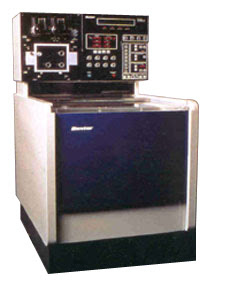
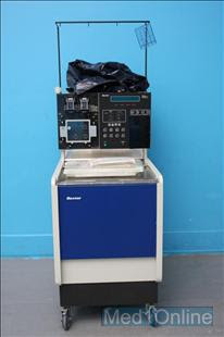
PLATELET REDUCTION USING BAXTER CS3000+

Platelet reduction procedure is very similar with collection procedure. Beware of the differences.


Kit : Standard Kit

Plate : Platelet TNX

Volume Target : 7000 mls

ACDA Ratio : 11 to 1 ml

Blood Test : Full Blood Count Pre and Post Procedure

PROCEDURE:

1. Select Procedure 1

2. Install and prime the kit

3. Connect patient

4. Press start to commence procedure

5. Check ACDA rate

6. Set the baseline after first spill over

7. When target is reached, press menu followed by start/resume to rinse back

8. Press halt/irrigate to finish procedure

9. Disconnect patient

Thursday 5 February 2009

DONOR PLATELETS COLLECTION USING BAXTER CS 3000+


Kit : Long Life Closed Circuit Kit
Plate : Platelet TNX
Volume Target : 5000 mls
ACDA Ratio : Between 9:1 ml to 10 to 1 ml
Blood Test : Full Blood Count Pre and Post Procedure
Target Yield : 5.5 x 10¹¹

PROCEDURE:
1. Select Procedure 1
2. Install and prime the kit
3. Connect patient
4. Press start to commence procedure
5. Check ACDA rate
6. Set the baseline after first spill over
7. When target is reached, press menu followed by start/resume to rinse back
8. Press halt/irrigate to finish procedure
9. Disconnect patient

Friday 3 October 2008

Panggilan Jiwa


I was working in Intensive Care Unit this morning doing plasma exchange for patient who has antiphosphatelipid syndrome with thrombocytopaenia. The aim of the procedure is to improve the platelets count. One of the British nurses was looking after the patient. I know from her thick accent. Anyway, she asked me to help her to give a patient a wash while the patient still in apheresis procedures. "No problem", I said, "I am a nurse, too". She did very good job, very thorough and very clean. I complement her by saying how good she was. She said to me, "I treat all the patients like how I treat my own family". I was so impress what she said. Sometimes we forget that the patient is a human being, too. Needs to be cared for, it is not because we have to, but because we want to. Some inner willing to help other people with a big heart. With a soul. In Indonesian we call it "Panggilan Jiwa"

Photo taken from All Saints, smh.com.au

Wednesday 1 October 2008

Com.Tec Fresenius Kabi part 2


It's about 6 months now that I have trialled Com.Tec Fresenius Kabi Machine. Three procedures I already mastered so far, Therapeutic Plasma Exchange (TPE), Stem Cell Collection (Auto MNC), and Immunoabsorption (TPE with column). Since I tried Spectra Optia already, all apheresis machines need to be close to that standard at least. This machine is not dissappointing at all. I thought TPE and Auto MNC procedure were ok. It's not until I tried Immunoabsorption procedure, I was blown away. It was very easy and very simple procedure, and also you can cut down the time of procedure, too. Marvelous. I would love to see how this machine perform with other procedures.
Up until now, I think I should have this machine at least one in my unit. How?

Thursday 3 April 2008

Trialling Com.Tec Fresenius Kabi


This month, we started trialling the new apheresis machine: Com.Tec from Fresenius Kabi, Australia. One thing is good about this machine is, able to do a few procedures including Therapeutic Plasma Excange and Stem Cell Collection. So far so good..... It seems like a combination between CS3000+ from Baxter (Fenwall) and Cobe Spectra from Gambro, BCT. The technology defenitely one step ahead of those machines. Very user friendly and easy to understand.... Might be later on I will be able to compare it with optia spectra from Gambro, BCT. I love optia....
With this machine, at the moment I only able to practice TPE procedure, may be in the next few days I will learn more about the stem cell collection... As a TGA auditor will come to our unit soon... it means we might put this issue on hold. Anyway, we have a few months to play with....

Friday 14 March 2008

TOPIK KEPERAWATAN KANKER

1. Refleksi arti dari keperawatan kanker
2. Biologi dari Kanker
3. Patogenesis Karsinogen
4. Peran dari sistem immunitas di dalam kanker
5. Prinsip dari terapi kanker(treatment modalitas):
a. Radiasi Onkologi
b. Obat-obatan anti kanker
c. Bioterapi dan terapi genetik
d. Transplantasi sel stem dan apheresis
6. Isu kesehatan dan keselamatan kerja untuk perawat kanker
7. Clinical Trial
8. Sindrom Paraneoplastik
9. Kegawat daruratan Onkologi
10. Efek dari kanker dan terapinya
a. Mual dan muntah
b. Nyeri pada kanker
c. Disfungsi sumsum tulang (bone marrow)
11. Isu perawatan suportif dan kualitas hidup
a. Qualitas hidup dan pengalaman hidup penderita kanker
b. Isu reproduksi dan sexualitas
c. Pendekatan dan memenuhi kebutuhan pasien yang akan meninggal
d. Spiritual dan kanker
12. Refleksi pengalaman penderita kanker

Friday 29 February 2008

JENIS DARI TRANSPLANTASI SEL STEM (SEL INDUK)

Sel Stem atau disebut juga sel induk adalah sel darah imatur yang dapat berproliferasi dan berdiffensiasi menjadi sel darah dewasa (matur), seperti sel darah merah, sel darah putih atau keping darah. Sel induk ini terbentuk di sumsum tulang (bone marrow). Sel stem yang berada di sumsum tulang dapat diambil secara langsung, maka area yang diambil biasanya berasal dari ileac crest. Proses ini harus dilakukan di kamar operasi. Sumsum tulang yang diambil berkisar 1,8 hingga 2,2 liter. Sumsum tulang ini terdiri dari campuran sel darah, plasma darah dan sel stem. Perawat Apheresis kemudian memisahkannya berdasarkan berat dari sel tersebut. Plasma darah adalah yang paling ringan , komponen ini tidak dipergunakan. Sel darah (biasanya adalah sel darah merah), diberikan kembali ke donor dan sel stem dipergunakan untuk transplantasi. Bila sel stem diambil dari darah tepi (peripheral), GCSF atau GMCF dipergunakan untuk menstimulasi sel stem bersikulasi di peredaran darah tepi.
Pertama-tama, kemoterapi dosis tinggi dengan atau tanpa radioterapi diberikan ke pasien untuk menghancurkan sumsum tulang. Kemudian sel stem diberikan sehingga sel stem yang baru dapat menolong menggantikan fungsi sumsum tulang yang lama yang sudah rusak.
Ada 3 jenis transplantasi sel stem:
1. Allogenik (Allograft) - berasal dari orang lain bukan dari pasien.
Donasi mungkin dari match atau mismatch sibling, keluarga dekat atau dari orang lain yang tidak ada hubungan darah yang disebut juga Matched Unrelated Donor (MUD). Dipergunakan untuk berbagai macam gangguan darah. Harus diberikan langsung (fresh) sebelum 72 jam. Bila tidak, harus dibekukan dengan cryopreservation setelah dilakukan kemoterapi pre transplant (conditioning).
Cord Blood, diambil dari ari-ari bayi yang baru lahir dan disimpan di Cord Blood Bank. Diberikan kepada pasien dari 2 ari-ari bayi yang berbeda (Double Cord Blood Transplant). Hitung CD34 positive pada cord blood biasanya kecil, jadi dibutuhkan 2 kantong.
Mini Allogenik, hampir sama dengan allogenik, tetapi penerima donor mendapatkan dosis kemoterapi yang lebih rendah (non-myeloblative) dengan atau tanpa radioterapi. Keuntungan metode ini, mengurangi efek samping dari kemoterapi sehingga pasien yang sebelumnya mungkin tidak dapat di transplantasi menjadi dapat, contohnya orang tua, dan pasien yang tidak pernah mendapatkan remisi. Suksesnya terapi ini bergantung pada mekanisme Graf Versus efek dari tumor (GVT).
2. Otologus (dari pasien sendiri)
Pasien mendonasikan sel stemnya sendiri yang kemudian diberikan kembali pada saat yang tepat sesudah kemoterapi dosis tinggi.
Indikasi: Tidak ada donor yang tersedia, tidak ada indikasi untuk mendapatkan allogenik.
3. Singenik
Transplantasi antara dua kembar identik.
(Dari berbagai sumber)

Tuesday 8 January 2008

Trialling Spectra Optia


Last week on 3rd and 4th of January 2008, I had a training with the new gambro Machine, Spectra Optia Apheresis System. Apparently we are the first one to try on in New South Wales. Very exciting.... It is incredible machine.... I really... really... love it and also very user friendly... and only a touch away to operate it..... As I am new in Apheresis world , this machine does help me alot how the system works. Unfortunately at this stage, this machine only able to do Therapeutic Plasma Exchange procedure. As Cobe Spectra machine will be discontinued in the next a few years, the replacement machine is so awesome... From the operator point of view, I will explain a little bit more about doing TPE with this little machine... Stay tune.

Tuesday 1 January 2008

Happy New Year 2008


Happy New Year 2008. It's been a long time that I haven't update my blog. One of my new year's resolutions is up dating my blog. Hopefully, I will up date my blog at least once a month. Finger cross.
Anyway... I was so happy that our unit achieved a TGA (Therapeutic Good and Administration) License last year. We are the first in NSW to have a license in Apheresis Unit and the 3rd in Australia. Mudah-mudahan saya dapat membahas sedikit mengenai TGA ini in the next few months.... I promise...
Hal lain yang saya harus diselesaikan di tahun 2008 ini adalah buku tentang Mieloma Multipel yang saya tulis bersama-sama dengan Eviana Tambunan dan Rindawani Simarmata. Mudah-mudahan tahun baru ini membawa semangat baru dalam diri kita masing-masing sehingga kita dapat memberikan sesuatu yang bermanfaat terhadap perkembangan keperawatan. Bravo....

Sunday 17 June 2007

ACUTE LYMPHOBLASTIC LEUKEMIA

QUESTION 1
Select a patient in your clinical setting and provide a brief descriptive profile outlining their age, diagnosis, disease stage and general treatment plan.
WT is a seventeen years old boy who was referred to Emergency Department by a GP with two weeks of legs petechia rash, hematuria, bilateral lower leg pain and general lethargic. It was also noted that he had a bruising and bleeding easily over last few days, tachycardic, and haematomesis. There were no epistaxis or melena presents.
From blood test showed that his blood counts were abnormal.
Haemoglobin: 110 l
White Cell Count: 268.5 c
Neotrhophills: 21.6 h
Platelets: 17 c
Circulating Blast Cells: 198.5 c
Lymp: 35.6 h
Mono: 8.9 h
RBC: 3.77 l
HCT: 0.33 l
Microscopy MSU:
WBC: >100 x 10,000,000
RBC: >100 x 6
Epi: <10
Chemistry: Positive of ketones, protein,
From blood test’s result, Tom was suspected with Acute Leukemia. Tom was then referred to haematology ward for further investigations after stayed overnight at children’s ward. These investigations include Bone Marrow Aspirate, CT Abdoment, Chest Xray, HIV V2V HSU serology and routine blood test such as FBC, EUC, CRP, LFT, Co agulation, & C3 nephritic factor.
After investigations were performed, Tom was finally diagnosed with Acute Lymphacytic Leukemia (ALL). The treatment plan for him includes:
To commence induction chemotherapy by using BFM 95 protocol.
Tissue typing of the family if allogenic stem cells transplant required in the future.
Sperm Banking.
Further Bone Marrow Biopsy and other test to view the response to induction therapy.
To understand leukemia, it is important to understand the process of blood formation. The formation of blood cells started in bone marrow which called haematopoiesis. Leukemia is a type of blood cancer affecting the bone marrow. Bone marrow is the spongy center of the bone that produces blood cells. All mature blood cells are originated from a pluripotent stem cell which is produced in bone marrow. Stem cell can be divided into two lineages, myeloid lineage and lymphoid lineage. These are a type of mature myeloid and lymphoid cells, including white cells or leukocytes (help the body fight against infection and other disease), red blood cells or erythrocytes (carry oxygen from the lungs to the body’s tissues and take carbon dioxide from the tissue back to the lungs), and Platelets (help form blood clots that control bleeding). These cells are normally produced in an orderly controlled way when the body needs them, but with leukemia that process gets out of control.

Leukemia means white blood. In leukemia the marrow produces too many immature white blood cells, which is called blasts. These immature white blood cells are abnormal shaped and cannot carry out their normal duties. These blasts are multiplying and crowding the bone marrow, as they interfere the production of other type of blood cells. Red cells, platelets and mature white cells will be affected. When they circulate in the body, they can be stayed in different places causing swelling or pain. Leukemia has a sudden onset and rapid progression of symptoms and disease.
Acute leukemia can be classified into acute myeloid leukemia (AML) and acute lymphoblastic leukemia (ALL). AML develops when maturation and proliferation of myeloid stem cell are abnormal, and ALL is established when abnormalities in maturation and proliferation of the lymphoid stem cells are occurred. In other word lymphocytic leukemia that affects the white blood cells called lymphocytes, which is the overproduction of the immature lymphocytes. Lymphocytes control the body’s immune response by finding and destroying foreign substances.

ALL is common occurred in children by two third (Cremer et al. 2002, 6). As the abnormal white blood cells cannot help the body fights against infection, frequent infections and flu-like symptoms such as fever and chills will occur. As the abnormal cells keep multiplying and move out into the body, they are tend to be collected in the lymph nodes organs such as liver or spleen or joints.

QUESTION 2
Outline the role of diagnosis and staging in the evaluation of your patient’s disease and its relevance to the general treatment plan.
Untreated ALL shows the presence of abnormal white blood count, haematocrit, haemoglobin, platelets counts, bone marrow (more than 5% blast) as well as the presents of signs and symptoms of the disease. The diagnosis can be made by performing full physical examination, blood test, bone marrow aspirate/biopsy and other additional tests. The physical examination is to find out the swelling in the liver, the spleen, and the lymph nodes under the arms, groin and the neck. From Tom’s physical examination were found that he had swelling under the arms, groin and the neck. He also had an enlarged spleen.

Blood test is performed to see what the cells look like under the microscope and compare the number of abnormal cell with the normal cells. Tom’s blood test showed that he had excessive white blood cells count (268.5) and circulating blast cells (198.5). There is no blasts should be circulating in the blood.

Bone marrow aspirate is a procedure by taking a sample of bone marrow using needle inserted into large bone, usually hip. Sometimes a small cykindrical piece of bone and marrow are also needed, this procedure is called bone marrow biopsy. This procedure will confirm weather or not the leukemia is present. The ALL can be established when 25% more of nucleated cells in the marrow contain lymphoblast.
There is no clear cut staging system for ALL according to Pui and Evans (1998, 2). They also noted that the diagnosis of ALL is depending on immunophenotyping. Unlikely AML which can be identified by the presence of auer rods, myelophoblastic leukemia, or monocyte associated esterases, ALL is lack of specific morphologic or cytochemical features (Pui & Evans, 1998, 2).
World Health Organisation (WHO) classified the types of leukemia in respect to where the cell formation is disruptive and with prognosis of outcomes. Cytogenetics can confirm the diagnosis and favourable or unfavourable outcomes.
Leukemic cell characteristic such as morphological features cytocemistry, immunologic cell surface and biochemical markers and cytogenic characteristics are important when diagnosing leukemia. However cellular classification using French, American and British (FAB) classification is limited for ALL, it is still used. The FAB classification classified ALL into three morphology types including L1 T-cell and B-cell in children, L2 T-cell and B-cell in adult, and L3 B-cell (Burkitt cell types). L1 morphology occurred in 84% of children with ALL, and 80% of Adult ALL have L2 morphology.
Immunophenotyping is more relevant to ALL when makes diagnose or prognosis. Immunophenotyping can be studied by flow cytometry. Flow cytometry and the use of monoclonal antibodies allow detecting cell surface antigents. Antigens are identified by cluster differentiation (CD) number.
The treatment plan for Tom is to commence induction chemotherapy with Vincristine, Daunorubicin, and Asparaginase without methotraxate.

QUESTION 3
Considering your answer to question 2, review the current literature and rationalize the treatment regime your patient is currently receiving.
The course of therapy for ALL can be divided into three stages:
1. Induction therapy.
2. CNS propylaxis.
3. Post remission therapy.

The aim of induction chemotherapy therapy is to achieve complete remission. Complete remission is defined by restoration of normal haematopoesis including less than 5% of blast cells present on bone marrow and no circulating blast cells.
Front-line therapy for children with ALL is mostly similar in all developed countries including Australia.

Induction chemotherapy used for Tom included:
Prednison 60 grams/m2/day per oral from day 1 to day 29.
Vincristine 1.5 grams/m2 per intravenous on day 8, 15, 22, 29. Daunorubicin 30 milligrams /m2 per intravenous on day 8 & 15. Asparaginase 5000 units/m2 per intravenous on day 12, 15, 18, 21, 24, 27, 30, 33.
It is called BFM 95 protocol with modification. For his treatment, metotraxate was not used.
The side effect of Asparaginase is quite significant. For some patients, coagulopathies, pancreatitis and allergic reaction could occur. Therefore, a test dose of 100 units of asparaginase subcute is required 45 minutes prior to the therapy. APPT, PT, Fibronogen level and Pancreatic Lipase test should be taken prior to asparaginase. If fibrinogen level <1.0 g/l or Lipase level are rising, a specialist has to be contacted. When pancreatitis is present, no further dose is to be given (RNSH Hematology Department, 2001).
Gated heart pool scan cardiac echo needs to be considered to rule out ventricular dysfunction if further dose of daunorubicin required, as this chemotherapy is toxic to the heart at cumulative dose of 400 mg/m2. For high risk ALL, additional dose might be added on day 22 and 29 (RNSH Hematology Department, 2001).
After normal hematopoesis was restored post induction therapy which is called in remissio, patient will be a candidate for intensification (consolidation) therapy. The treatment will commence shortly after the induction of remission. According to RNS Haematology Protocol for Chemotherapy (2001), the treatment protocol for Tom is called BFM 95 Protocol MCA which includes Methotrexate given in high dose, 6-mercaptopurine per oral plus Cytarabine. For High risk ALL, Etoposide and Asparaginase are added without 6-Mercaptopurine. This protocol is called BFM 95 Element HR-3.
Pui et al (p. 7) also suggested that intensive multi drug therapy commenced post induction of remission will make remission lasting-longer, especially in young adults. Cyclosphomide and Cytarabine might be benefit for patient’s with T-cell ALL. It was also suggested that Cytarabine with dose will improve the outcome of patient with standard, risk, or high risk ALL (Pui et al. p. 7).
Some suggestion made by Gaynon (2002, 1) that the progress of the treatment also can be achieved by improving the post induction therapy. Pui et al (p. 7) concluded that the successful cure of ALL depends on the development of more effective multi drugs regime on well designed clinical trial. Tom has not started with intensification therapy yet as he just commenced with induction therapy.
Most of the patient with ALL can be expected to attain complete remission status following appropriate induction therapy. According to Pui et al. (p.6) the rate of complete remission for ALL in children now ranges from 97 to 99 percent and lower in adult from 75 to 90 percent. All patients with ALL are at risk of developing central nervous system involvement during the course of their disease.

QUESTION 4
Identify two actual or potential complications that may confront your patient in light of their treatment regime or disease progression.

Nausea and vomiting was perceived by patients to be the most distressing side effect of chemotherapy (Coulbourne, 1995, p. 442; Potter & Schafer, 1999, p.2). Chemotherapy drugs have been cited to often cause nausea and vomiting within 1-2 hours. However, some drugs induce these symptoms several hours after treatment. Symptoms observed ranged between a mild to severe level of severity, depending on the types of the drugs and the doses used. Usually this condition was cited to resolve within 24 hours or over several days (Held-Warmkessel, 1998, p. 43).
Chemotherapy drugs were seen to have a potential inducing nausea and vomiting in a variety of ways. Drugs associated with pronounced nausea and vomiting were noted to be that of Camustine, Cisplatin, Cyclophosphomide, Cytarabine, Dacarbazine, Dactinomycin, Doxorubicin HCl, Lomustine, Mechlorethamine HCL, and Procarbazine HCl (Glick, Griffith & Mortimer, 1998, p. 50). According to RNSH Chemotherapy Patient Information Leaflet (2001), Asparaginase and Daunorubicin have a potential nausea and vomiting side effects. It is not only on early side effects, but also on late side effects. However, mild nausea might be expected in Vincristine.
Dodd, Onishi, Dibble & Larson (1996, p. 160) determined the differences in the side effects of nausea, vomiting and retching between younger and older patients receiving chemotherapy. This research reported that younger patients’ consistently had higher nausea, vomiting, and retching than older patients. Therefore, this finding suggested that older patients could be given more aggressive chemotherapy regimes, as they were seen to be able to tolerate them better than younger patients (Dodd et al., 1996, p. 160).

Fatigue was reported as the most common and debilitating response to chemotherapy treatment (Carrol-Johnson et al, p. 116; Held-Warmkessel, 1998, p. 42; Mast, 1998; Richardson, et al. 1998). Patients receiving chemotherapy for the treatment of cancer, frequently experience severe fatigue, which was seen to be highly distruptive and distressing (Richardson & Ream, 1997, p. 35). Fatigue will be expected for patients who are receiving Asparaginase, Vincristine, and Daunorubicin (RNSH Chemotherapy Patient Information Leaflet, 2001).
Fatigue was also cited to increase over the time that the patient receives chemotherapy treatment (Berger, 1998, p. 51-62; Carrol-Johnson, et al. 1998, p 116; Richardson, et al. 1998) and after treatment (Carrol-Johnson, et al. 1998, p 116). The level of fatigue was reported to occur frequently in the afternoon and early evening, and perpetuate throughout the day (Richardson, et al. 1998). It was cited that fatigue reverted to baseline levels before the next chemotherapy cycle, and persisted for years after treatment has been completed (Carrol-Johnson, et al. 1998, p 116).
Fatigue was cited to affect the general well-being of patients, by weakness, and negative effects such as their general well-being, their specific body parts, change in self image and their daily activities. There were reported to be varying degrees and dimensions of ‘being tired’. Tiredness was not only seen as physical in nature, but also mentally exhausting (Messias, Yeager, Dibble & Dood, 1997, p. 45). Medication, nutritional status, work, and other physical conditions were seen to implicate the cause of fatigue. Patients, who were viewed to have more energy, were reported to be less tired with a lower dose of chemotherapy or on the termination of treatment (Messias et al. 1997, p.46).
When it came to patients’ awareness and expectations, some persons were reported to not have prepared for the degree or the extent of fatigue that they may experience, while others recognized the invetability and the expectations of the fatigue on the bases of their prior knowledge (Messias et al. 1997, p.46). It was also reported that "patient express frustration, worry, concern, displeasure, fear, depression, and disappointment regarding their fatigue" (Messias et al. 1997, p.46). Some patients experience frustration by the extent and the dimension of their tiredness. Others reported being annoyed or bothered by the fatigue. Fatigue was also viewed to be associated with mood and energy levels. It was seen to diminish patients’ levels of functioning and slow their pace of operation, decrease their threshold for activity, and make the person totally disabled. Patients who were tired have been seen to have difficulty with sleeping, a need for increased sleep, and also observed to lack a desire to be active. As a result of the adverse reactions from chemotherapy, patients who had fatigue were cited to be working fewer hours, taking increased time off, experiencing diminished interest and productivity, and frequently expressing their concern about the possible negative effects of not working or not going to school (Messias et al. 1997, p.46).

Dimensions of fatigue were described as the extent of fatigue, distress caused by fatigue, the influence of fatigue on the ability to engage in social activities and impact of fatigue on work related activities. By knowing these dimensions of fatigue, the nurse will be able to provide more effective help for individual who experienced fatigue (Richardson et al. 1998).

Monday 16 April 2007

PHERIPHERAL VENOUS CANNULATION AND VENEPUNCTURE WORKSHEET

Ini adalah worksheet peripheral venous cannulation and venepuncture course dari Sydney South West Area Health Service yang saya ikuti dulu. Mudah-mudahan bermanfaat bagi anda yang akan mengikuti course yang sejenis.

1. List the three layers found in Arteries and Veins?
Tunica interna
Tunica media
Tunic externa

2. Circle which vessel contains valves?
Arteries Veins

3. Describe the purpose of valves, which are found in above vessel.
The valves prevent back flow and in this way aid the flow of blood returning to the heart.

4. Please label the diagram below:
Maaf diagramnya tidak ada. Jawabannya dimulai dari

Sebelah kiri:
* Cephalic Vein

* Cephalic Vein
* Median Cubital Vein

* Median Vein
* Basilic Vein

Sebelah Kanan:
* Median Veins - Cephalic
* Cubital Vein
* Basilic Vein

* Basilic Vein

* Cephalic Vein

* Metacarpal Vein
* Dorsal Vein

5. What are the indication for performing IV cannulation?
For IV therapy and IV medication.

6. List the indication for performing venepuncture.
For various blood test.

7. What are the five legal consideration when performing venepuncture &/or cannulation procedure?
Verbal consent from patient
Medical order
Confidential documentation duty of care
Compliance with Hospital policy.
Compliance with procedural guidelines.

8. Explain the five basic rules for reducing the risk of infection when performing venepuncture &/or cannulation.
Correct hand washing technique.
Correct IV site preparation.
Maintenance of aseptic technique.
Correct choice of site.
Secure cannula firmly in place.

9. What protective equipment is used by the nurse when performing venepuncture &/or cannulation?
A protective eyewear, gloves, kidney dish, protect IV cannula/vacutaneous system and sharp bin.

10. Describe how you would identify your patient prior to the procedure of venepuncture &/or cannulation?
Ask patient her/his full name, date of birth, medical record number against both armband and clinical record such as notes, label (venepuncture)

11. List the nursing considerations when positioning a tourniquet on a patient’s limb for venepuncture &/or cannulation?
Ensure tourniquet is functional, sate and clean.
Do not have the tourniquet on too tight or for too long
Use tourniquet approx 5-10 cm above the venous access site.

12. What is the maximum tourniquet application time and state the reason?
3 minutes. Will cut the circulation to lower site.

13. Selection of a vein for IV cannulation / venepuncture depends upon.
1. Patient’s age
2. Reason for insertion
3. Size of cannula
4. Condition of vein
5. Expected duration
6. Patient co-operation
7. Position of vein
8. Solution/ fluid ordered
9. Required floe rate

14. List four sites to avoid when selecting a cannulation /venepuncture site.
1 . Bruised, tender or phlebitis areas
2. Hardened or sclerotic veins
3. Flexion areas
4 . Veins in lower limbs

15. List the most appropriate cannulation site
Cephalic vein
Metacarpal vein
Basilic vein

16. List the contraindications for performing IV cannulation.
No patient consent
No doctor order
Patient who have special medical conditions listed in hospital policy such as burns, children, infected skin.

17. What safety aspects of the cannulation / venepuncture equipment should the nurse check prior to use?
Ensure the equipments are intact and undamaged/
Ensure within the expiry date.

18. What are the four (4) complication associated with IV cannulation and venepuncture?
Haematoma.
Infiltration.
Phlebitis.
Thrombophlebitis.

19. Complete the following sentence.
The skin site is cleaned with alcohol wipe (chlorhexidine) and allowed to dry for 30 second to prevent infection and irritation when performing cannulation.

20. How many unsuccessful IV cannulation/venepuncture attempts can you have on any one patient?
Cannulation two times venepuncture two times

21. You have just inserted the IV cannula, and you notice a haematoma has formed. What action do you take?
Remove cannula, apply pressure to the site at least two minutes, monitor the site and document it.

22. When inserting the cannula, how do you ensure it will not cause a sharps injury. Give your rationale.
Ensure sharp bin inclose range, always use kidney dish when removing the sharp/ needle out. Pull back until click is heard. Always using needle lock, therefore there is no naked needle left.

23. What documentation is required when cannulation has been undertaken?
Cannulation size, site used, along with any other associated patient management provided.

24. The patient has complaint of pain and swelling around the site 36 hours after insertion of the cannula. What action do you take?
Remove cannula, note the site for pain or swelling or other sign of infection. Document the findings.

25. What safe labelling procedure should you follow when labelling the blood tubes?
Before and after taking the blood (venepuncture), patient’s name, medical report number, date of birth and test/s required must be checked with patient (family/staff mamber if it unable).

26. List the equipment you will need to prepare for a venepuncture proceure.
Alcohol swabs
Disposable gloves
Vacutainer
Tourniquet
Vacutainer needle + barrel
Needle (s)
Biohazard bag (s)
Evacuated specimen tube (s)
Gauze/wool swabs
IV pressure/ bandaid/ macropore
Pads sharp bin

27. In preparing the venepuncture needle system, what safety aspect should the RN/EN on the needle system?
Ensure equipment intact and not damage. Needle are sealed and check expiry date.

28. How many times can a vacutainer be engaged before the vacuum is lost?
Once

29. Identify the order in which blood specimens should be collected when collecting several specimens from a single venepuncture?
Number 1,2,3,4 &5 in order of collection. Type of Tube.
5 Addictive tube (green, grey, mauve, yellow, pink)
1 Blood cultures
3 Coagulation tube (blue)
2 No addictive tube
4 Serum tube with gel separator (white)

30. If drawing blood from a cannula that is newly inserted or from an arterial line… what volume of blood should you discard before collecting the first blood specimen?
Nil

31. If collecting blood via butterfly needle and there is citrate in the blood tube…how much blood do you discard first to remove the dead space in the butterfly tubing?
½ mls if 21 G needle used.

32. Circle the correct procedure for venipuncture. (A B or C)
A. Removed the vacutainer tube from needle, release the tourniquet, remove the needle and apply direct pressure to the needle site.
B. Release the tourniquet, remove the needle with the engaged vacutainer tube, apply digital pressure to the vessel above the needle site before removing the needle.
C. Release the tourniquet, remove the vacutainer tube from the needle, apply digital pressure to the vessel above the needle site before removing the needle, removing the needle and apply direct pressure to the needle site.

Sunday 25 March 2007

Therapeutik Apheresis: Overview


Prosedur Therapeutik Apheresis:
Adalah prosedur dimana komponen dari darah dikeluarkan dari tubuh dengan maksud mengurangi jumlah sel yang diinginkan atau juga menghilangkan sel mediator dari penyakit.
Rasionalnya adalah prosedur ini jauh lebih efektif mengeluarkan pathogen yang berada di dalam darah yang mana berkonstribusi terhadap penyakit daripada tubuh sendiri mampu mengeluarkannya.


Prosedur Therapeutik Apheresis terdiri dari:
1. Therapeutik Plasma Exchange (TPE)
2. Red Blood Cell Exchange (RBCE)
3. Cellular Depletion
4. Immunoadsorption
5. Photopheresis
6. LDL Apheresis


TPE
Mengeluarkan plasma dalam jumlah besar dari tubuh dan menggantikannya dengan cairan yang sesuai.
Indikasi: TTP, GBS, MG, Sindrom Good pastureSelain itu prosedur ini juga mengeluarkan mediator penyakit yang berada di dalam plasma, seperti:
Antibodi, antigen, abnormal plasma protein, cholesterol, produk-produk sampah metabolik, obat-obatan atau racun
.


RBCE
Adalah tindakan mengeluarkan atau membuang sel darah merah dan menggantikannya dengan sel darah merah dari donor yang normal.
Indikasi: Sickle cell disease, Thalassemia, Malaria, Babesiosis.


Cellular Depletion
Adalah tindakan mengeluarkan abnormal sel yang meningkat didalam darah secara pathologis. Berguna untuk menurunkan resiko sehubungan dengan vaskular statis.
Indikasi: Leukositosis (Sel darah putih >100 000/mcL)
Thrombositosis (sel trombosit/platelets >500 000
Erythrositosis (hematocrit >60%)


Immunoabsorpsi
Adalah tindakan mengeluarkan sel mediator dari penyakit yang berada didalam plasma dan mengembalikan kembali plasma yang sudah ditherapi ke pasien.
Indikasi: ITP, Rhematoid arthritis, refraktori platelets dan sindrom hemolitik uremik.


LDL Apheresis
Adalah tindakan mengeluarkan kolesterol LDL dari plasma melalui dua kali proses apheresis.
Indikasi: Hiperkolesterol (kolesterol LDL > 300 gr/dL atau > 200 gr/dL dan simtomatik).


Photopheresis
Adalah tindakan memodulasi aktifitas dari lymphosit dengan menggunakan teknologi apheresis, obat-obatan dan sinar ultraviolet.
Indikasi: Cutanous T Cell Lymphoma (CTCL), Graft Versus Host Disease (GVHD), penolakan pada transplantasi ginjal akut atau kronik serta Phemphigus vulgaris.


ELEMEN DALAM PROSEDUR APHERESIS


1. Pemisahan komponen darah
Centrifugal Force memisahkan sel berdasarkan gravity dari sel tersebut.
a> Plasma (paling atas).
b> Buffy coat (ditengah - thrombosit, lymphosit, monosit dan granulosit).
c> Sel darah merah.


2. Akses vaskularisasi
a> Antecubital
b> femoral catheter
c> Subclavian Catheter
d> Jugular Access
e> Port
f> Arteriovenous Fistula atau Graft


3. Antikoagulasi, ada 2 macam antikoagulasi yang dapat dipakai:
a> ACDA (Acid Citrate Dextrose - Formula A)
Menyatu dengan kalsium didalam darah sehingga proses pembekuan darah tidak terjadi, dari IX ke IXa, dari X ke Xa dan dari prothrombin ke Thrombin.
Akibatnya dapat menurunkan pH darah, mencegah terjadinya pembekuan darah pada thrombosit.
Sangat cepat dimetaboliskan oleh tubuh.
Dapat menyebabkan hipokalsium.


b> Heparin
Mencegah terjadinya formasi thrombus sehingga thrombin tidak dapat diproduksi yang berguna dalam pembentukan fibrin dari fibrinogen. Tidak ada fibrin, pembekuan darah tidak terjadi.
Sangat lambat dimetabolisme oleh tubuh.
Bekerja secara sistematik.
Dapat menyebabkan thrombocytopenia.

4. Cairan pengganti, setiap prosedur mempunyai cairan pengganti yang berbeda.
a> TPE, ada beberapa cairan yang bisa dipakai, tetapi yang lebih sering dipakai adalah 4% Albumin.
- Kristaloid (tidak mengandung protein).
Contoh: 0.9% Sodium Chloride.
- Koloid (mengandung protein).
Contoh: 4% Albumin, Fresh Frozen Plasma (FFP) atau Cryopersepitate.


b> RBC
Cairan penggantinya adalah sel darah merah.


c> Cellular Depletion
0.9% Sodium Chloride atau 4% Albumin.


d> Collum Prosedur (secondary plasma processing)
Tidak membutuhkan cairan pengganti.


5. Manajemen cairan yang seimbang
a> Isovolemia cairan yang diambil sama dengan cairan pengganti
b> Hipovolemia cairan yang diambil lebih besar daripada cairan pengganti
c> Hipervolemia cairan yang diambil lebih rendah daripada cairan pengganti


6. Frekuensi tindakan
a> Pada penyakit akut
Progres penyakit cepat
Membutuhkan tindakan yang sering (setiap 1 hingga 2 hari atau setiap 12 jam)


b> Pada penyakit kronik
Progres penyakit lambat
Tidak membutuhkan tindakan yang sering (setiap minggu atau bulan)

7. Potensi efek samping - preventif, gejala dan tindakan
a> Potensi terjadinya efek samping diantaranya adalah:
Hipokalsemia
Ketidak seimbangan elektrolit tubuh
Hipotensi
Syncope vasovagal
Reaksi alergi
TRALI (transfussion related acute lung injury), komplikasi yang dapat mengakibatkan kematian sesudah mendapatkan transfusi darah.


b> Pencegahan terjadinya efek samping
Sebelum memulai prosedur sebaiknya:
Mengkaji pasien secara komprehensif, diantaranya diagnosa, riwayat kesehatan, obat- obatan dan hasil laboratorium.
Mengkorekasi ketidak seimbangan elektrolit tubuh bila terjadi.


c> Gejala dan Tindakan.
Hipokalsemia
Tanda dan gejala
Perasaan baal dan kesemutan
Kedinginan
Fibrasi pada dinding dada
Tetani
Aritmia pada jantung hingga serangan jantung


Pencegahan
Cek kalsium dan elektrolit dalam darah
Berikan kalsium dan elektrolit dengan tepat sesuai dengan kebutuhan.
Pergunakan penghangat transfusi darah.


Tindakan
Hentikan/tunda prosedur hingga pasien merasa lebih baik
Turunkan kecepatan "inlet pump"
Berikan kalsium secara intravenous
Ketidak seimbangan eletrolit lain yang mempunyai tanda dan gejala yang hampir sama dengan hipokalsemia, tetapi pemberian kalsium tidak menghilangkan gejala, diantaranya Hipomagnesium, hipokalemia, alkalosis metabolik.


Hipotensive
Gejalanya:

Sakit kepala
Meningkatnya kecepatan nadi
Pernafasan pendek
Berkeringat.


Pencegahan
Naikkan persentasi albumin.
Berikan cairan tambahan
Pilih hasil cairan akhir, positive (hipervolemia), bila prosedurnya TPE


Tindakan
Tunda prosedur
Posisi kaki lebih tinggi dari kepala
Berikan cairan tambahan


Vasovagal syncope
Gejala:
Aprehensi
Pusing
Mual
Kecepatan nadi menurun.
Hipotensi
Berkeringat


Pencegahan
Komunikasi dengan pasien
Jelaskan ke pasien tentang prosedur sehingga pasien dapat mengerti apa yang terjadi
Alihkan perhatian pasien.


Tindakan:
Tunda prosedur
Posisi kaki lebih tinggu dari kepala
Berikan cairan tambahan
Tindakan hampir sama dengan hipotensi.
Reaksi Alergi


Gejalanya:
Gatal
Kemerahan pada kulit
Bengkak-bengkak
Sulit bernafas


Pencegahan
Cek apakah pasien mempunyai alergi
Berikan obat anti alergi seperti antihistamin


Tindakan
Hentikan prosedur
Kontak dokter
Berikan obat-obatan sesuai dengan yang diorder.


TRALI
Gejalanya:
Oedema pada paru-paru yang bukan diakibatkan oleh jantung
Dyspnea
Sianosis
Hipotensi.
Demam
Menggigil


Tindakan:
Seperti pada pertolongan respiratory, termasuk bantuan ventilator.


Sumber:

Successions 2: Spectra System Therapeutic Reference Book. Gambro BCT.